
Standing MRI
We’re pleased to be able to offer MRI investigation of lameness problems.
When a horse is admitted for MRI, we allow them a night of hospitalisation to settle into our hospital environment. A more settled horse means we will obtain higher quality images with less sedation.
On the day of the MRI, the horse has an intravenous catheter placed to allow sedation to be given easily.
The horse’s shoes are removed and radiographs taken to ensure no metal fragments remain in the hoof, because metal may render the images as non-diagnostic.
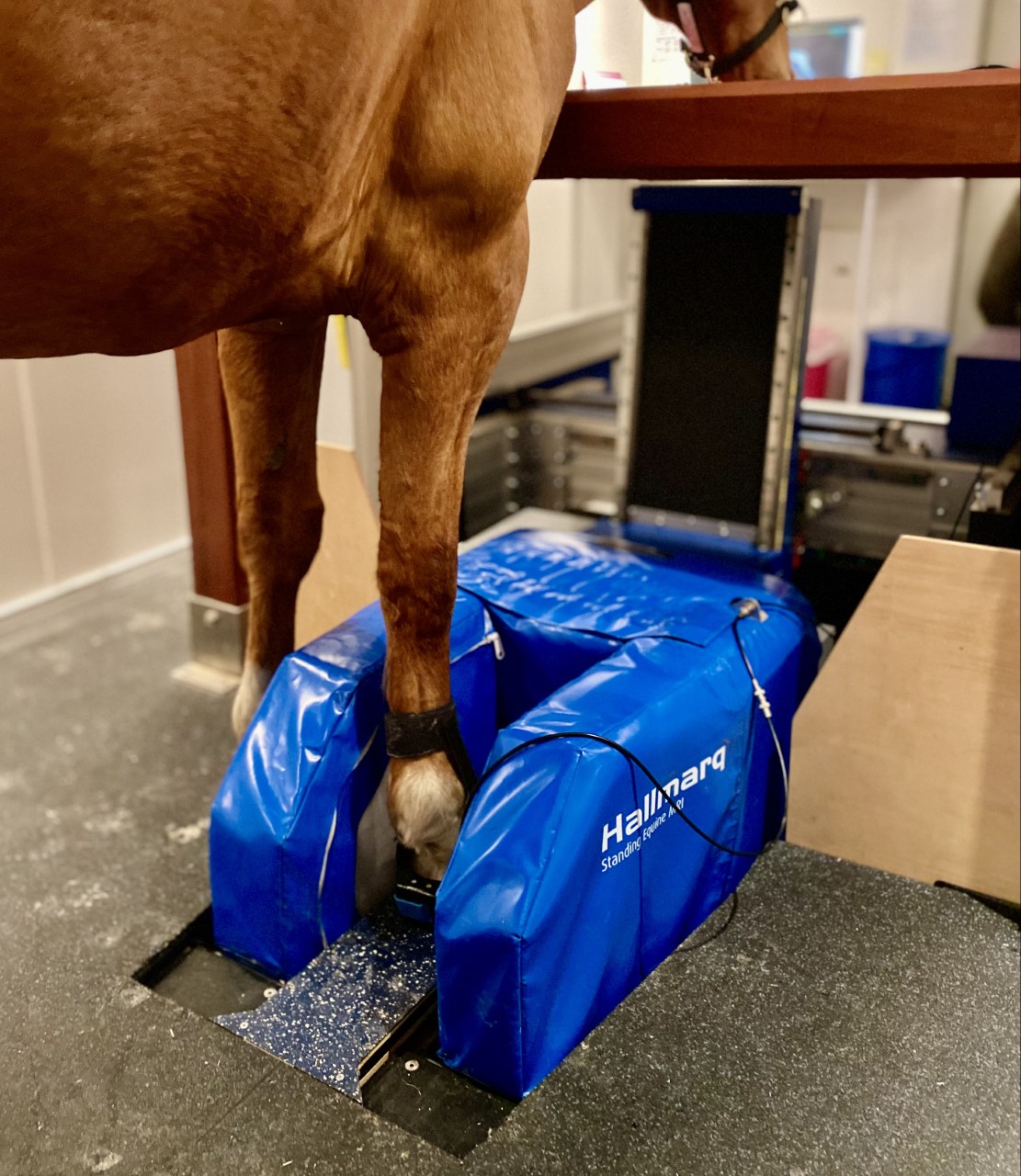
After this preparation, the horse enters the MRI suite. The scans are taken with the horse standing and the affected limb positioned in a large U-shaped magnet.
The images are obtained over a one to three hour period, depending on which area is to be scanned, after which they are sent electronically to an image interpreting specialist.
We usually expect the report from the consultant to be ready within 72 hours, at which point your vet will then be able to discuss the results and formulate the most appropriate treatment plan with you.
About the procedure
If your horse has been booked in for an MRI scan, we ask that you bring your horse into the hospital prior to scanning, usually the day before, to allow your horse to settle in.
This allows the horse to become accustomed to our hospital environment and means we can use lower doses of sedation. Less sedation provides us with less movement blur and better quality images.
Please bring your passport, so that we can check your horse’s vaccination status and that Section IX (exclusion from the food chain) has been signed.
We have all the feedstuffs your horse will require; however, if your horse has any allergies, please bring your own food as appropriate.
Please bring rugs and any medications or supplements that you would like your horse to have during their stay.
To facilitate repeat sedation doses, a catheter will be placed into the vein. It is standard practice to clip and aseptically prepare the area prior to catheter insertion. If you have concerns regarding this, please discuss it with a member of our team.
The appropriate shoes will need to be removed before scanning, i.e for forelimb scans both front shoes will be removed and for hindlimb scans both hind shoes will be removed.
After shoe removal, radiographs will be taken to ensure there are no small pieces of metal remaining in the foot; if there is, we will remove these prior to scanning.
The scan time will be determined by the areas to be imaged and the demeanour of your horse in our scanning environment.
After scanning, your horse will be taken back to a stable, monitored post sedation and fed when appropriate.
Once the scan is completed, the images are sent to our selected specialist for interpretation, who will endeavour to report their findings to us within 72 hours.
If your horse returns home on the same day, please ensure water is readily available and soft wet feedstuffs are given, as prolonged sedation can result in decreased bowel motility and increased colic risk.
These precautions minimise the colic risk; however, we advise monitoring your horse closely for the following 12 to 24 hours, especially with regard to faeces passed.


